baby chest x ray exposure
With dental x-rays there is hardly any exposure to any part of the body except the teeth. Your doctor may use x-rays to help place tubes or other devices in your body or to treat disease.

Interpretation Of Neonatal Chest Radiography
A protective lead apron to shield certain parts of the body.

. Full legfull spine imaging is performed at 180 cm using CR. Erect chest X-rays are taken at 180 cm. This brochure is to help you understand the issues concerning x-ray exposure during pregnancy.
There are also differences in exposure factors between the two countries. However these dose levels arent used in diagnostic imaging. Chest X-rays require very little preparation on the part of the person getting it.
Lateral cervical spines are taken at 150 cm. Arms are not superimposed over lateral chest wall this can mimic pleural thickening. When X-rays of the ribs the state of the bone mechanism is visualized and the spine can be partially seen.
Radiation exposure from X-rays may slightly raise the risk of later cancer especially in children who have had many tests with high radiation exposure. Full legfull spine imaging is performed at 180 cm using CR. Most researchers agree that babies who receive a small dose of radiation equal to 500 chest x -rays or less at any time during pregnancy do not have an increased risk for birth defects.
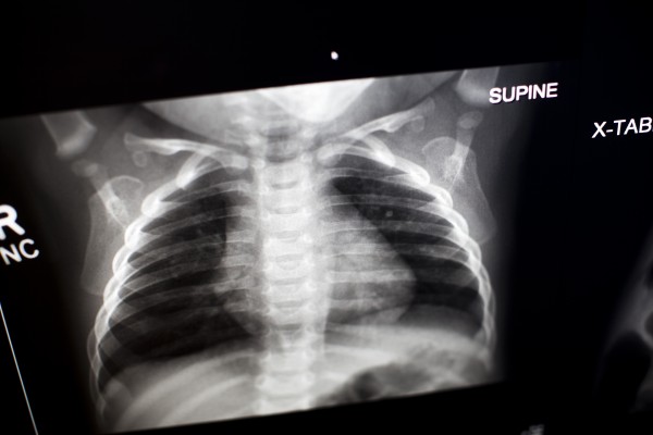
Most neonatal chest X-rays are AP films unless the baby is made to lie prone Lucency of soft tissue shadow - darker the soft tissue more. The chest X-ray is the most frequently ordered radiological. The entire lung fields should be visible from the apices down to the lateral costophrenic angles.
These measurements were repeated for seven X-ray machines available in. For example the amount of exposure to the fetus from a two-view chest x-ray of the mother is only 000007 rad. Canada uses lower kV Figure 1 but Norway uses lower mAs Figure 2.
Although X-rays are still occasionally over or under exposed a discussion of penetration now best serves as a reminder to check behind the heart. The publication of this study and exposure chart could act. Rotation Soft tissue bone Thymus -.
The chin should not be superimposing any structures. The most sensitive time period for central nervous system teratogenesis is between. Doctors agree that exposure to the small amount of radiation produced during an.
Because they spin around the body taking multiple images CT scans can deliver radiation doses that are up to 200 times higher than an average chest X. The degree of ionizing radiation is not considered dangerous to human health so X-rays can be considered a good alternative to ultrasound 1 computed. In addition to radiation exposure Burstein points out theres a high association between receiving a chest X-ray for bronchiolitis and antibiotic prescribing which is.
The risk of harm to your baby depends on your babys gestational age and the amount of radiation exposure. Exposure to extremely high-dose radiation in the first two weeks after conception might result in a miscarriage. The left hemidiaphragm should be visible to the edge of the spine.
Exposure factors X-ray tube voltage kVp tube current mA exposure time s related to every view was set on the X-ray units and then measurements were performed. All distal extremity exposures are taken at 110115 cm SID. X-ray exams provide valuable information about your health and help your doctor make an accurate diagnosis.
X-ray examinations on the arms legs or chest do not expose your reproductive organs to the direct beam. Exposure to high-dose radiation two to eight weeks after conception might. Long-term problems are very small.
Diagnostic x-rays can give the doctor important and even life-saving information about a. All distal extremity exposures are taken at 110115 cm SID. As a benchmark for other medical imaging departments and to promote discussion on digital X.
A well penetrated chest X-ray is one where the vertebrae are just visible behind the heart. Lateral cervical spines are taken at 150 cm. However x-rays of the torso such as the abdomen stomach pelvis lower back and kidneys have a greater chance of exposure to the uterus.
A chest X-ray is a painless noninvasive procedure with few risks. Erect chest X-rays are taken at 180 cm. The only increased risk to these babies is a slightly higher chance of having cancer later in life less than 2 higher than the.
X-rays use a small amount of radiation about the same levels that occur naturally in the environment. Exposure 527 Neonatal Chest X-ray. At Stanford we take extra precautions to minimize our patients exposure to radiation including using.
35 cm x 43 cm or 43 cm x 35 cm. The median exposure parameters in Canada for mobile neonatal chest imaging are 60 kV at 15 mAs with an inherent filtration of 20 mm Al Figure 3. Plain X-ray shows the existing damage to the internal organs and the whole chest.
Often smaller mini detectors are used for the neonate chest x-ray. See Safety in X-ray Interventional Radiology and Nuclear Medicine Procedures for more information. Radiation exposure from X-rays does not pose any short-term problems.

Pediatric Chest X Ray In Covid 19 Infection European Journal Of Radiology

Reliability Of Chest Radiograph Interpretation For Pulmonary Tuberculosis In The Screening Of Childhood Tb Contacts And Migrant Children In The Uk Clinical Radiology

X Ray For Kids Children S Health Orange County

Neonatal Pneumonia Radiology Reference Article Radiopaedia Org

Indications For Chest X Rays In Children And How To Obtain And Interpret Them

What Is An X Ray For Kids Radiology And Medical Imaging

X Rays And Unshielded Infants Raise Alarms The New York Times

Neonate Chest Supine View Radiology Reference Article Radiopaedia Org

Diagnostics Free Full Text A Pictorial Review Of The Role Of Imaging In The Detection Management Histopathological Correlations And Complications Of Covid 19 Pneumonia Html

Interpretation Of Neonatal Chest Radiography

Neonatal Chest Radiography Influence Of Standard Clinical Protocols And Radiographic Equipment On Pathology Visibility And Radiation Dose Using A Neonatal Chest Phantom Radiography

Neonatal Pneumonia Radiology Reference Article Radiopaedia Org

Pediatric Chest Pa Erect View Radiology Reference Article Radiopaedia Org

Interpretation Of Neonatal Chest Radiography

Interpretation Of Neonatal Chest Radiography

Pathogens Free Full Text Chest Imaging For Pulmonary Tb Mdash An Update Html
Simple Diagnostic Model For Pneumonia In Kids To Reduce Need For X Rays Imaging Technology News

Pediatric Chest X Ray In Covid 19 Infection European Journal Of Radiology
